Optomap Monaco Retinal Eye Exam
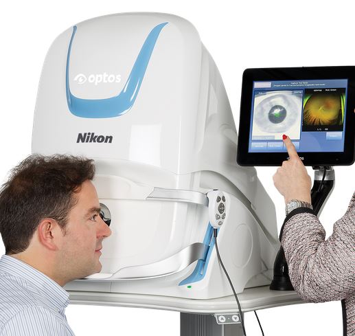
Optos Monaco provides Ulta-Widefield Retinal Image. 200 degrees of Retina in zero point five seconds - 0.5sec! What does that mean to you? That means the doctor can get a beautiful, panoramic image of your retina really, really quick. Potentially alleviate the need for dialation. AND, it provides a baseline image to work with going forward as your eyes age. Its OK, all of our eyes age, you are not alone! The Optos Monaco assists our doctors to detect common ocular pathologies such as diabetic retinopathy, hypertensive retinopathy, high cholesterol.
This is the latest, state of the art, Retinal eye exam technology that we have been waiting for!
While traditional Optos provides only wide field retinal imaging, the Optos Monaco is the first device that combines retinal wide field images with OCT or Optical Coherence tomography. The OCT integration is a diagnostic tool that uses light waves to capture high resolution cross-section images of the inner layers of the retina. The analogy is similar to an ultrasound or a CT scan for the eye and allows us to see deeper into the retina, which is something we can’t normally do even during dilation. Our doctors are now able to detect ocular pathology such as macular degeneration and diabetic retinopathy earlier in hopes of preventing larger ocular issues down the road. In just 90 seconds, the Monaco can quickly capture images in color, autofluorescence and OCT using a multi-modality feature. This new OCT integration has changed the way our doctors manage various ocular conditions.
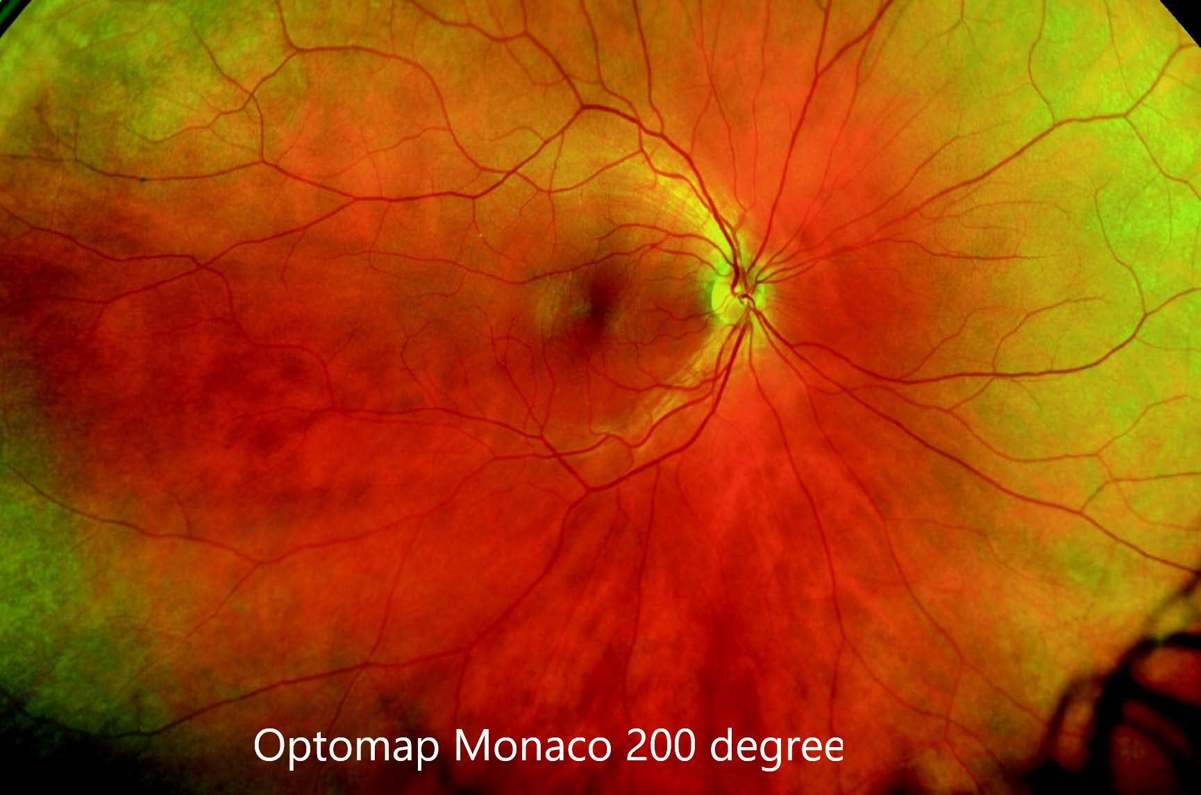
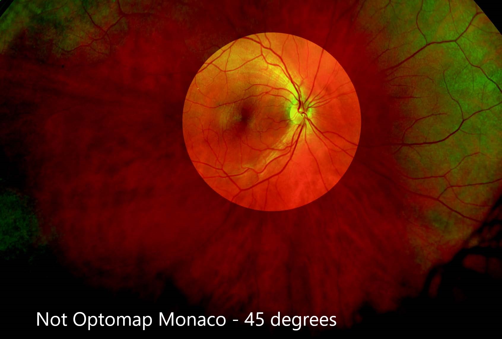
Click here to read more about OCT or Optical Coherence Tomography here.With traditional, small-field, and even widefield retinal imaging, only 10-100 degrees of the retina can be captured. Optomap is clinically validated for ultra-widefield retinal imaging that can capture 82% or 200 degrees of the retina. With optomap auto-montage, up to 97% or 220 degrees of the retina can be imaged.
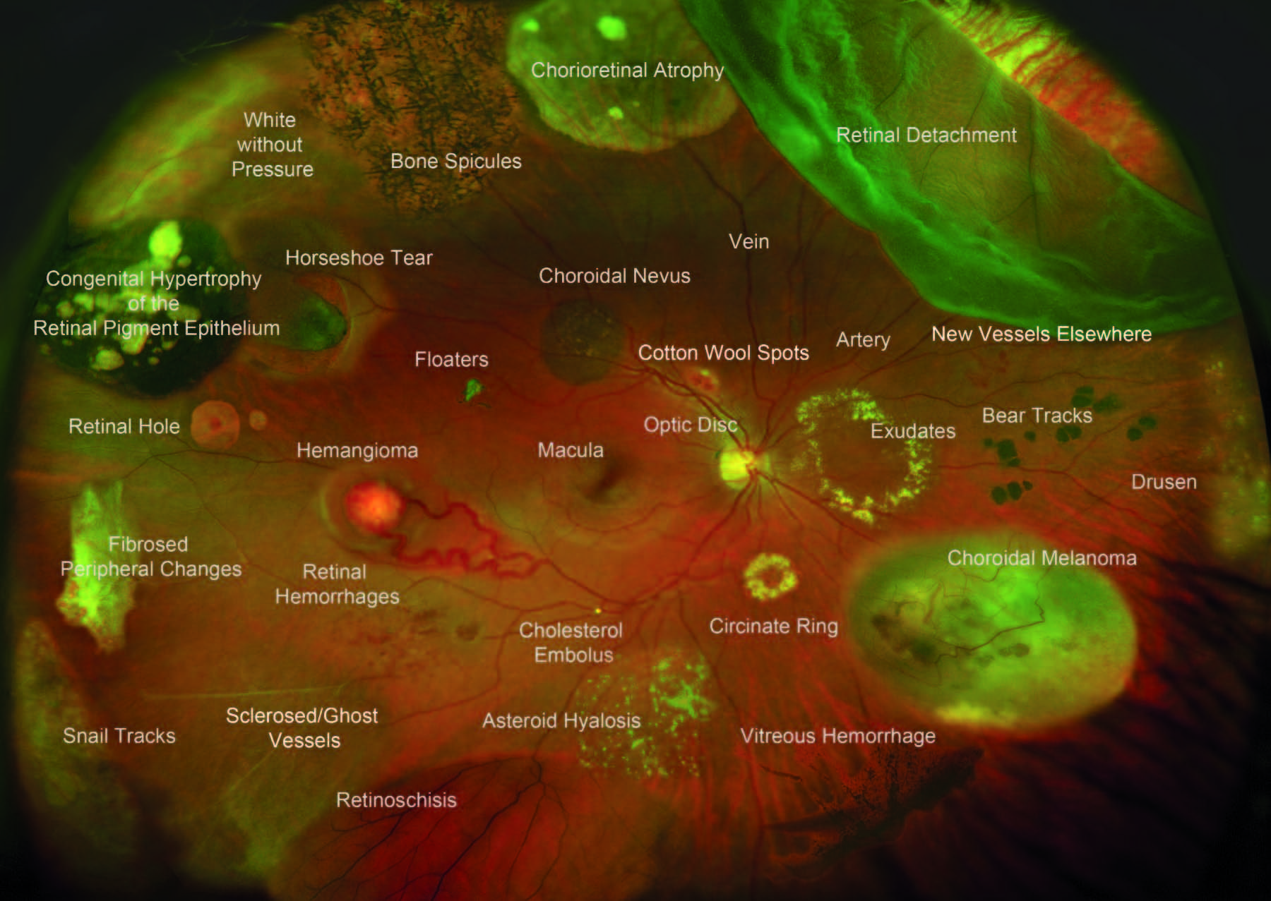
Optomap offers unparalleled views of the retina which provide:
Wide Field Retinal Imaging

© 2024 Fine Eyewear & Eyecare - Higher Quality. Better Vision. ™